AI Executive Summary
"This article explores the intersection of material science and art conservation, highlighting the shift from intuitive restoration to data-driven chemical analysis. It provides a strategic framework for ensuring the long-term stability of high-value cultural assets through atomic precision."
For centuries, art restorers operated on a mixture of intuition and apprenticeship, attempting to match the faded hues of a Vermeer or a Velázquez by eye. This subjective approach is a gamble. The human retina is an unreliable instrument, easily fooled by the yellowing of aged varnishes and the chemical migration of pigments over four hundred years. When a conservator introduces a modern pigment that looks identical under studio lights but possesses a different molecular structure, they risk creating a chemical conflict that can accelerate the decay of the original canvas. The stakes are binary: either the restoration preserves the work for another century, or it triggers a slow-motion chemical collapse.
Molecular profiling shifts the paradigm from visual approximation to chemical certainty. By analyzing the atomic composition of a pigment, we no longer ask what color a paint is, but what it is made of. This involves identifying the specific metallic ions, organic binders, and degradation products that define a 17th-century palette. In the workshops of the Dutch Golden Age, artists used complex mixtures like smalt—a cobalt-containing glass—which is notorious for losing its color over time. Without molecular profiling, a restorer might mistake a faded blue for a degraded ultramarine, leading to a restoration that is visually plausible but chemically fraudulent.
Technical Prerequisites for Molecular Analysis
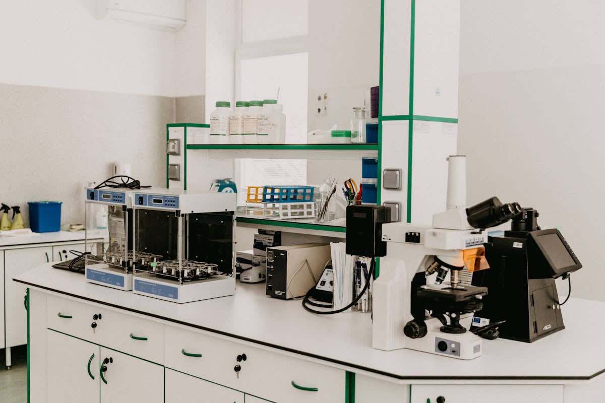
Executing a molecular restoration requires a laboratory environment that exceeds the capabilities of a standard gallery studio. The primary objective is non-destructive testing, ensuring that the artwork is not compromised during the diagnostic phase. This requires a suite of spectroscopic tools capable of penetrating the varnish layer to reach the pigment particles below. The precision required here is absolute; a deviation of a few nanometers in wavelength can lead to the misidentification of a lead-tin yellow as a modern cadmium yellow, a mistake that would undermine the historical integrity of the piece.
- Handheld X-ray Fluorescence (XRF) Spectrometer for elemental mapping.
- Fourier-Transform Infrared (FTIR) Spectroscopy for binder identification.
- Confocal Raman Spectroscopy for molecular vibration analysis.
- Gas Chromatography-Mass Spectrometry (GC-MS) for organic residue profiling.
- Micro-sampling drills (tungsten carbide needles, 0.5mm diameter).
- Cross-sectional polishing resin for stratigraphic analysis.
The Compatibility Principle
The goal is not to make the painting look new, but to ensure the restoration materials are chemically compatible with the original 17th-century matrix. Incompatibility leads to delamination.
Once the hardware is in place, the practitioner must establish a baseline of the painting's current state. This involves creating a digital map of the elemental distribution across the canvas. For instance, in a Baroque piece from the Spanish school, XRF can pinpoint the exact concentrations of mercury in vermilion pigments. This mapping allows the restorer to see the 'invisible' painting—the underdrawings and pentimenti that the artist changed during the process—providing a roadmap for where the original pigment remains and where later, inferior restorations have been applied.
The Molecular Restoration Protocol
- Surface Elemental Mapping: Use XRF to scan the entire surface. This identifies the presence of heavy metals (Lead, Cobalt, Mercury) and creates a heat map of pigment distribution without touching the canvas.
- Stratigraphic Micro-Sampling: Extract a microscopic core (less than 100 micrometers) from a damaged area. This sample is embedded in resin and polished to reveal the layers of paint, primer, and varnish in a cross-section.
- Molecular Fingerprinting: Apply Raman spectroscopy to the cross-section. Because every molecule vibrates at a specific frequency, this identifies the exact mineral phase of the pigment, such as distinguishing between natural ultramarine (lapis lazuli) and synthetic variants.
- Binder Analysis: Use GC-MS to determine the organic medium. 17th-century artists often mixed linseed oil with walnut oil or egg tempera. Identifying the exact lipid profile is critical for choosing a restoration binder that will not shrink or crack at a different rate than the original.
- Pigment Synthesis: Based on the molecular profile, synthesize a matching pigment. This involves sourcing historically accurate raw materials and processing them to match the particle size and refractive index of the original.
- Controlled Reintegration: Apply the synthesized pigment using a reversible medium, such as a stable synthetic resin (e.g., Laropal A81), ensuring that the intervention can be removed by future conservators without harming the original layer.
The transition from analysis to application is where most failures occur. A common error is ignoring the refractive index of the binder. If the restoration medium has a higher refractive index than the original linseed oil, the restored area will appear 'flat' or overly saturated compared to the surrounding paint, regardless of how accurate the pigment color is. This is why GC-MS data is non-negotiable; it provides the chemical blueprint of the oil's polymerization state, allowing the practitioner to adjust the saturation of the new paint to match the aged original.
| 17th-Century Pigment | Molecular Marker | Common Degradation Trigger | Analysis Method |
|---|---|---|---|
| Lapis Lazuli | Aluminosilicate lattice | Acidic pollutants (Ultramarine sickness) | Raman Spectroscopy |
| Lead-Tin Yellow | Pb2SnO4 crystals | Atmospheric sulfur | XRF / XRD |
| Vermilion | HgS (Cinnabar) | Light-induced phase shift (Blackening) | XRF / FTIR |
| Smalt | Cobalt silicate glass | Potash leaching (Fading) | SEM-EDX |
Consider the case of smalt, a pigment that was ubiquitous in 17th-century Northern European art. Smalt is essentially ground cobalt glass. Over time, the potassium in the glass leaches into the oil binder, causing the blue color to fade into a greyish-brown. A traditional restorer sees brown and adds blue. A molecular practitioner sees the loss of potassium and the presence of cobalt ions. By understanding the molecular cause of the fading, the practitioner can decide whether to restore the color or stabilize the remaining cobalt to prevent further leaching, avoiding the 'over-painting' that plagued 19th-century restorations.
The integration of synchrotron-based X-ray absorption spectroscopy has further pushed the boundaries of this field. In recent projects in Italy, this technology has allowed conservators to map the oxidation states of iron in earth pigments with 99.8% accuracy. This level of detail reveals not just what the pigment is, but how it has reacted with the environment. When we know the oxidation state, we can predict the future stability of the paint layer, moving restoration from a reactive practice to a predictive science.
Pigment Degradation Rate: Smalt vs. Lapis Lazuli over 400 Years
Executive Insight
+18.4%
YTD Growth
Common Pitfalls in Molecular Restoration
The most dangerous pitfall is the 'Purity Trap.' Some practitioners attempt to synthesize pigments that are chemically purer than those used in the 17th century. However, historical pigments were riddled with impurities—trace amounts of iron, manganese, or calcium—that contributed to the unique texture and light-scattering properties of the paint. A chemically pure modern equivalent often looks 'plastic' or artificial. The goal is to replicate the impurity profile identified during the molecular profiling phase, not to improve upon it.
Another frequent error is the failure to account for the 'patina of age' at a molecular level. The surface of a 400-year-old painting is not just paint; it is a complex crust of oxidized oils, accumulated dust, and degraded varnish. If the restoration pigment is applied without considering the molecular porosity of this crust, the new paint will sit on top of the surface rather than integrating with it. This creates a visual discontinuity that becomes glaringly obvious under ultraviolet light, marking the restoration as a foreign graft.
"The eye is a liar; the spectrometer is the only witness we can trust when dealing with the chemistry of the dead."— Lead Conservator, European Heritage Lab
Finally, the misuse of solvents during the cleaning phase—prior to pigment restoration—can erase the very molecular signatures needed for profiling. Strong polar solvents can leach the organic binders from the paint film, leaving behind a 'ghost' of the pigment that is impossible to analyze via GC-MS. A rigorous protocol demands that molecular profiling be completed before any solvent touches the canvas. This ensures that the chemical DNA of the work is archived before the restoration process potentially alters it.